X-Ray For Braces? Why Are They Needed?
Last Updated on July 4, 2023 by Gio Greenard
When it comes to getting braces, X-rays are an important part of the process. Even though many people know what to expect when getting metal braces, not everyone is aware of what to expect when getting an X-ray. X-rays are a crucial part of the orthodontic treatment process and can help orthodontists create a personalized treatment plan for a healthy, straight smile. A dental X-ray can also provide you with valuable information about your treatment plan based on the information gathered.
While braces can seem like a ho-hum boring piece of the realm of teenagers, an orthodontist must make sure that they will be helpful to the customer. This is where x-rays come in handy.
FAQ
What are Orthodontic X-Rays?
X-rays can be a frustrating thing to take when your child is small. If they won’t sit still for the dentist, why would they for the orthodontist when they take x-rays? However, the orthodontic x-rays serve a different purpose than they would via a dentist’s office. The main type of X-ray used is called a bitewing. This type gives the taker a detailed picture of a small section of teeth.
Lighter areas on the X-ray indicate:
- Enamel
- Fillings
Darker areas indicate:
- Root canals
- Decay
- Bones.
The orthodontists are trained to read the light and dark areas in order to determine if there are issues in a particular area of the mouth. Normal tissues look different than abnormal tissues on the X-rays and knowing the difference could help figure out what to be done next.
Some reasons an X-ray would be taken include:
- A first time at the office
- To see the bones if pain is reported
- To see how recovery is going after a surgery
Why would an x-ray be taken?
Among the reasons orthodontists take x-rays, to begin with, is to be able to identify abnormalities. Whether these are tumors or pathogens that could end up doing serious harm, many show up on x-rays. Often times, these issues are first identified on the x-rays – whether they are done in Manhattan Beach or in Paris. After they have been identified, the patient is referred for a treatment based on what is wrong.
Another reason that x-rays will be taken when having an orthodontic visit is to check on the progress of the treatment. Braces do move the teeth and straighten them out – and for some people, this causes their roots to shorten. This is incredibly rare since only 2% of patients experience this. However, the X-rays can help decide if there is a problem with having too many teeth in the mouth. Sometimes the treatment will begin and teeth will begin to align, but then an overcrowding problem arises. If this happens, a decision is reached about what to do – whether that is the tooth is pulled or if the teeth need to be shifted in a different direction.
What is The Issue
If the overcrowding issue is the problem, an X-ray is usually taken before treatment so that a proper treatment can be done. One or two will be taken along the way if braces are the decision – but most patients only have an X-ray taken before and after the treatment.
A fourth reason an X-ray would be taken is to determine if the jaw bone has any issues that need to be fixed. Whether this means that the jaw is too big, too small, or off center, it can be a problem. When the issue has been determined, tooth position is looked at. If there was no jaw issue, the teeth are still looked at. Missing teeth, extra teeth, or even misplaced teeth are of particular interest to orthodontists. All the information in the X-ray is used to determine if a surgery or tooth extraction is needed to correct the problem.
Kids
While children may not sit still for long, getting them to sit still for an X-ray is incredibly useful. These X-rays are used to determine growth patterns in children. They are also used to look at any adult teeth that have yet to push through. Predictions of what kind of growth can be made via these X-rays as well.
Adults
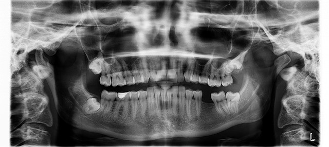
In adults, panoramic X-rays (or X-rays that show the entire mouth like a book) are used to identify if a particular treatment will work. Results can be quantified and can be predicted to a certain degree. Changes with treatments can also be seen via the panoramic X-rays.
Lateral cephalograms (or side view X-rays) are used for the same reasons. Like the bitewing, these offer a look at a small section of teeth. Unlike the bitewing, however, the lateral cephalograms show little detail on any one tooth – and show the side of the skull and spinal cord. Computer tracing can be used to help determine how the teeth look with the lips and nose – drawn in of course. Teeth can be traced and a triangle drawn to figure out if there are reasons to use a specific treatment.
While getting an X-ray at the doctor’s entails heavy coats and a large room, the same technology used by orthodontists has regulations set by the state. There is no need to worry about unnecessary exposure. The operators are hand-picked to be sanctioned by the state. The room that the equipment is in has also been sanctioned before it was ever introduced to the X-ray machine.
There is More
On another note, the machines expose a person to less radiation than they’re exposed to in an airplane. The “as little as reasonably achievable” – or ALARA – The rule of thumb helps to keep this practice safe. The idea is to keep the patient exposed to as little radiation as possible. Most people have between three and five X-rays in three to four years, which means it is safe to expect one a year – two if needed for treatment.
Moreover, since the X-rays are stored digitally now, they can be transferred to any office via email. There’s virtually no need for a patient to receive another X-ray if there is little time between the time of the X-ray and the change in offices.
All in all, the X-rays taken by any orthodontist will be used to safely determine what kind of treatment is needed. Should a patient refuse to take an X-ray, a treatment may not be able to be started until more is known about the state of their teeth, gums, jaw, bones, etc. It’s possible this can only be done through X-rays – depending on what is in need of assessment at that time.
Conclusion
When it comes to getting braces, X-rays are an important part of the process. X-rays are a crucial part of the orthodontic treatment process and can help orthodontists create a personalized treatment plan for a healthy, straight smile. A dental X-ray can also provide you with valuable information about your treatment plan based on the information gathered.

Dr Patti Panucci attended the University of Louisville School of Dentistry for four years, where she graduated with a DMD degree (May 2000) among the Top 10 in her class. Following that, she headed west to Los Angeles to complete her three-year residency at one of the top-ranked orthodontic programs in the country – the University of Southern California.
Along with her certificate in orthodontics, Dr. Panucci earned a master’s degree in craniofacial biology. During those three years, she fell in love with Southern California beach life and decided that this was where her future lay.


Leave a Reply
Want to join the discussion?Feel free to contribute!